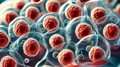
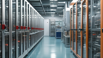
Cell culture research supports vaccine development, biologics manufacturing, cancer studies, and regenerative medicine. Its importance is expanding rapidly. Grand View Research estimated the global cell culture market at approximately USD 22.7 billion in 2023, with continued growth expected through 2030. The report links this expansion to pharmaceutical investment, bioprocessing demand, and advances in three-dimensional models.
Yet growth does not guarantee reliable results. A cloudy flask, an unexpected pH shift, or a single unnoticed contamination event can compromise weeks of work. R. Ian Freshney, a leading cell-culture educator and author, wrote, “Cell culture is both an art and a science.” That observation remains practical. Skilled researchers read cell morphology, track passage history, and question results that appear unusually convenient. They also recognize that no protocol is flawless.
This article, “10 Tips for Successful Cell Culture Research,” introduces habits that protect consistency from thawing to data analysis. It considers aseptic technique, authentication, mycoplasma testing, media preparation, confluence control, and transparent record keeping. The International Society for Cell and Gene Therapy and major suppliers, including ATCC, continue to stress quality control and traceability across laboratory workflows. These recommendations are not substitutes for institutional training or validated procedures. They are a working foundation.
Small details matter. Labeling a flask before opening the incubator can prevent confusion later. Recording the exact passage number can reveal hidden bias. Some failures remain difficult to explain. That is uncomfortable, but useful. Reliable cell culture research grows through careful repetition, honest documentation, and a willingness to challenge attractive results.
10 Tips for Successful Cell Culture Research
Cell culture begins with a research objective, not a flask. Define the biological question, measurable endpoint, cell model, and decision criteria before starting. The model should match the tissue, disease, or pathway under study. Record passage number, seeding density, medium changes, incubation conditions, and morphological observations. Small details become evidence later.
A reliable workflow includes aseptic technique, authenticated cell sources, contamination screening, controlled thawing, and consistent handling time. Mycoplasma testing deserves special attention; contamination can remain invisible while altering growth, metabolism, and gene expression. The World Health Organization’s Laboratory Biosafety Manual, fourth edition, stresses risk assessment, validated procedures, and documented training. These are fundamentals, not administrative decoration. Nature’s 2016 survey of 1,576 researchers found that more than 70% had failed to reproduce another scientist’s experiments. That figure should make every protocol feel less permanent.
Use biological replicates, technical replicates, appropriate controls, and predefined exclusion rules. Separate exploratory work from confirmatory experiments. Keep raw images and instrument files, not only polished figures. A 2021 report from the National Academies emphasized transparent reporting as a foundation for reproducible science. Still, perfect consistency is unrealistic. I have seen healthy-looking cultures produce unstable results after minor handling changes. That failure is useful, if recorded honestly. Review the objective when the cells behave differently, rather than forcing the data to fit the hypothesis.

Cell Culture Fundamentals and Research Objectives
The chart presents typical planning intervals for essential cell culture activities. Daily observation and documentation help detect contamination or abnormal morphology early, while scheduled medium changes, passaging, mycoplasma testing, and cell-line authentication support reproducible research. Exact intervals should always be adapted to the cell type, medium, growth rate, and laboratory standard operating procedures.
Successful Cell Culture Research: Laboratory Setup, Equipment, and Safe Aseptic Practices
A reliable culture begins with a controlled laboratory layout. Keep clean supplies separate from waste, and restrict unnecessary movement near the work area. Use a certified Class II biological safety cabinet for open manipulations. Check airflow certification at least annually, following CDC and NIH BMBL guidance. Wipe surfaces before and after work with a validated disinfectant. Allow the required contact time. Shortcuts here create invisible risks.
Wear dedicated coats, gloves, and eye protection. Change gloves after touching phones, doors, or paperwork. Inspect pipettes regularly, and use filtered tips for routine transfers. Label every vessel with cell identity, passage number, date, and operator initials. Record incubator temperature, carbon dioxide, humidity, and alarm events each day. Keep backup cultures, but avoid overcrowding the incubator. WHO laboratory guidance emphasizes documented procedures, training, and risk assessment rather than equipment alone.
Cell-line authentication and mycoplasma testing should be scheduled, not postponed until results look strange. The Nature 2016 survey of 1,576 researchers reported that more than 70% had failed to reproduce another scientist’s experiment, highlighting the value of transparent records and controls. My own weak point is assuming a quiet incubator means a healthy culture. It does not. Observe morphology, growth rate, and media color consistently. Photograph unusual changes. Review failed runs without hiding them; imperfect technique often reveals the next improvement.
| No. | Research Tip | Laboratory Setup or Equipment | Recommended Practice | Control or Acceptance Point | Typical Frequency |
|---|---|---|---|---|---|
| 1 | Design a clean, well-organized workspace | Separate cell culture activities from corridors, food areas, chemical storage, and high-traffic work zones. Use smooth, non-porous, easy-to-disinfect surfaces. | Keep only necessary materials inside the work area. Arrange supplies so clean items move into the workspace and waste moves out without crossing paths. | Work surfaces are uncluttered, visibly clean, and free from unnecessary paper, cardboard, and personal items. | Before and after each session |
| 2 | Use a certified biological safety cabinet correctly | Use a suitable Class II biological safety cabinet for procedures that may generate aerosols or involve potentially hazardous biological materials. | Allow the cabinet to run for the period specified in the facility procedure before work. Keep front and rear grilles unobstructed and avoid rapid arm movements. | Cabinet certification is current, airflow alarms are normal, and materials do not block the intake or exhaust grilles. | Certification at least annually and after relocation or major service |
| 3 | Maintain stable incubation conditions | Use an incubator appropriate for the cell type, commonly controlled near 37 °C with humidified air and approximately 5% carbon dioxide for bicarbonate-buffered media. | Minimize door openings, avoid overloading shelves, and allow the chamber to recover after loading. Use an independent temperature or carbon dioxide check when required by the laboratory procedure. | Temperature, carbon dioxide, humidity, and water-pan condition remain within the validated laboratory range. | Record daily or according to the validated monitoring plan |
| 4 | Choose and monitor the correct culture vessel | Select treated or untreated cultureware according to whether the cells are adherent or suspension-based. Use vented closures or other validated gas-exchange designs. | Label each vessel with cell line, passage number, medium, date, operator initials, and any relevant treatment information. | Labels remain legible, complete, and securely attached throughout the culture period. | At setup and during every media or passage change |
| 5 | Apply disciplined aseptic technique | Provide appropriate laboratory coats, gloves, eye protection, disinfectant, sterile pipettes, and a dedicated container for contaminated waste. | Disinfect gloves before entering the cabinet, work from clean to dirty areas, keep containers closed, and never place sterile tips or bottle caps on the work surface. | No direct contact occurs between sterile surfaces and non-sterile objects; spills and exposure events are reported promptly. | Every manipulation |
| 6 | Control media quality and storage | Store basal media, supplements, and reagents under their specified conditions. Use sterile, labeled aliquots when repeated handling could increase contamination or degradation risk. | Check expiry dates, appearance, lot information, and storage history. Warm only the volume needed for immediate use and avoid repeated temperature cycling. | Media show no unexpected turbidity, particles, color change, or precipitate, and are within the approved use period. | Before use and during routine inventory checks |
| 7 | Monitor cell morphology and growth | Use an inverted microscope with suitable illumination and a calibrated imaging or counting method when quantitative measurements are needed. | Inspect cultures for confluence, attachment, shape, debris, and signs of stress. Record observations consistently using predefined categories or images. | Culture appearance matches the expected profile for the cell type and experimental stage; unusual changes trigger review. | At each observation or media-change event |
| 8 | Passage cells at a consistent stage | Use a validated detachment method for adherent cells or a controlled dilution and centrifugation procedure for suspension cells. | Maintain consistent seeding density, passage interval, and recovery time. Avoid excessive confluence, prolonged enzymatic exposure, and unnecessary mechanical stress. | Viability, attachment, doubling behavior, and morphology remain within the laboratory’s established ranges. | At every passage |
| 9 | Prevent and detect contamination | Maintain dedicated incubator space where possible, use sealed secondary containers for transport, and provide validated procedures for decontamination and waste disposal. | Inspect cultures for turbidity, unexpected particles, rapid pH shifts, fungal structures, or abnormal growth. Test for mycoplasma using an approved method at defined intervals and before critical studies. | Suspect cultures are isolated, clearly marked, and not used until the investigation is complete. | Visual checks each session; mycoplasma testing periodically and after suspected exposure |
| 10 | Document, authenticate, and work safely | Use controlled records for passage history, cell source, storage location, equipment checks, deviations, and experimental conditions. Maintain appropriate cryogenic storage and chemical safety controls. | Authenticate important cell stocks, preserve low-passage master stocks, follow approved biosafety procedures, and dispose of biological and chemical waste using the correct streams. | Records are complete and traceable; only approved, identified cultures are used; incidents and deviations are documented. | At every experiment, stock transfer, and incident |
Selecting the right cells begins with the research question, not convenience. Use authenticated cells with documented passage history, origin, and genetic background. Keep passage numbers low when possible. High passage cells may drift in morphology, growth rate, or gene expression. A 2023 laboratory-quality report identified misidentification and contamination as persistent threats to reproducibility. Check morphology daily. Record changes.
Media selection deserves equal care. Match nutrient requirements to the cell type, then verify pH, osmolality, and serum dependence. Small formulation changes can alter attachment and differentiation. Prepare media with traceable lot records, and avoid repeated freeze-thaw cycles.
A 2024 industry market analysis estimated the global cell-culture sector above 20 billion dollars, reflecting rapid expansion but not guaranteed consistency. Market growth does not replace validation.
Control the physical environment precisely. Use the recommended temperature, carbon dioxide level, humidity, and seeding density. Overcrowded flasks often produce uneven nutrient access and stressed cells. Sparse cultures may attach poorly. Monitor confluence rather than relying only on calendar timing. Include negative contamination checks and quarantine questionable cultures. Follow good laboratory practice guidance, including complete records of operators, dates, deviations, and corrective actions. My own weak point has been assuming stable growth means healthy biology. It does not. Replicate critical conditions, compare independent cell lots, and document results before changing multiple variables.
Standardizing Cell Handling, Passaging, and Experimental Procedures
Reliable cell culture begins with a written standard operating procedure. Define vessel type, seeding density, medium volume, incubation settings, and passage limits. Keep these details visible near the work area.
Use consistent aseptic technique every time. Disinfect surfaces, organize materials before opening containers, and minimize exposure to the surrounding air. Check cell morphology daily under the microscope. Record attachment, confluence, debris, and unexpected color changes. Small details matter.
Thaw cells promptly and allow recovery before demanding experiments. Confirm identity and routine contamination status through approved laboratory procedures. Replace cultures at defined passage ranges, rather than relying on memory. Passage at a consistent confluence.
Overgrown cultures can change their behavior. Under-seeded cultures may grow unevenly. Prepare medium with the same warming practice and timing. Sudden temperature changes can stress cells.
Use matched controls, replicate wells, and clearly labeled vessels. Record operator, date, passage number, and any deviation from the protocol.
Our early records were too vague, making comparisons difficult. That mistake still informs our training.
When results look unusual, review handling records before changing the experimental design. Standardization is not rigid perfection. It is a practical way to recognize variation, correct weak steps, and protect the credibility of each result.
Reliable cell culture begins with disciplined observation, not hurried media changes. Check morphology, attachment, confluence, and color during each scheduled inspection. Record the date, operator, passage number, vessel, medium, and incubator readings. A quick photograph can reveal gradual changes that notes miss. Keep raw observations, even when they seem insignificant. Small clues matter.
When a culture looks stressed, compare today’s record with its previous two entries. Sparse attachment may reflect seeding density, handling damage, or unsuitable timing. Unexpected turbidity requires isolation and an approved contamination assessment. Do not rescue questionable material by mixing it with healthy cultures. Confirm temperature, carbon dioxide, humidity, and water levels before changing several variables. One change at a time makes troubleshooting clearer. It is slower, but more defensible.
I once focused on cell shape and overlooked a steady fall in growth rate. That mistake taught me to track doubling time and viable cell counts consistently. Use predefined acceptance limits, and document every deviation from the protocol. Review trends weekly with another trained researcher when possible. Records should explain what happened, what was checked, and why a decision was made. Some problems remain uncertain. State that uncertainty plainly.